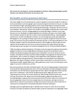
Find all the latest content on bifurcation lesions published on this website. The percutaneous treatment of coronary bifurcation lesions is very dynamic with an ongoing evolution in clinical approaches and techniques and the management of more complex cases continuing to evolve. Keep up-to-date about all these changes along with the latest research and devices as well as advances in imaging.
Image is adapted from classification and treatment of coronary artery bifurcation lesions: putting the Medina classification to the test. Diagnosis Angiography. Coronary artery bifurcation is detected on angiography. Shown below are an animated and a static angiogrpahy images depicting bifurcation of a left coronary artery lesion. Encircled in.There are multiple classification systems, which can be used to characterize bifurcation lesions (Fig. 22.2).One of the more complex systems is the Lefevre system (), which attempts to characterize the lesions by the location of the stenosis and angulation of the lesion (Fig. 22.3).Although descriptive, it is rather cumbersome to use for day-to-day practice and consensus societies such as the.However, true bifurcation lesions are Medina 1,1,1, 1,0,1 and 0,1,1 lesions, which are all summarized as B2 lesions when using the Movahed classification. This letter discusses this important issue.
Update on disease: percutaneous coronary intervention of bifurcation lesions Review Coronary artery bifurcation lesions pose a particular challenge in the field of interventional cardiology.
Coronary bifurcation lesions are frequently encountered in daily clinical practice by in-terventional cardiologists and account for 20% of all coronary lesions treated by percu-taneous coronary intervention (PCI) 1,2. Although clinical outcomes have been improved.
The accuracy and precision of quantitative coronary angiography (QCA) software dedi-cated for bifurcation lesions compared with conventional single-vessel analysis remains unknown. Furthermore, comparison of different bifurcation analysis algorithms has not been performed. Methods.
Although several classification systems of bifurcation lesions have been proposed to facilitate PCI, planning the treatment approach according to the angulation of the bifurcation and the degree of plaque burden seems to be most practical ().The Y-shaped lesion is defined when the SB and the main vessel (MV) angulation is less than 70%, while that of the T-shaped lesion is greater than 70%.
Medina classification of coronary bifurcation lesions: 1 is used to indicate the presence of stenosis and 0 the absence of stenosis in each of the three segments in order of importance: proximal main branch, distal main branch, and side branch.
FFR Guided Treatment Of Significant Bifurcation Lesions. Dr. Nicolas M. Van Mieghem obtained his medical degree and cardiology degree at the Catholic University of Leuven, Belgium.
The Medina classification is a straightforward system that has been popularly received. 8 However, prior randomized studies have included small to medium sample sizes and inadequately evaluated clinical outcomes according to angiographic distribution in patients with coronary bifurcation lesions, especially comparing true and non-true.
Role of lesion preparation in the treatment of bifurcation lesions. Case Review Session. Andrejs Erglis. Latvian Centre of Cardiology. Pauls Stradins Clinical University Hospital. Dr. Andrejs Erglis Role of lesion preparation in the treatment of bifurcation lesions. Case Review Session. Latvian Centre of Cardiology Pauls Stradins Clinical University Hospital Riga, Latvia. Lesion Preparation.
This banner text can have markup. web; books; video; audio; software; images; Toggle navigation.
Atherosclerosis is a narrowing of the arteries caused by a buildup of plaque. Arteries are the blood vessels that carry oxygen and nutrients from your heart to the rest of your body. As you get.
Relationship between intracranial internal carotid artery calcification and enlarged cerebral perivascular space Neuroradiology, May 2017 Xiao-Xiao Tao, Ge-Fei Li, Yi-Lan Wu, Yi-Sheng Liu, Ying Zhao, Yan-Hui Shi, Mei-Ting Zhuang, Tian-Yu Hou, Rong Zhao, Feng-Di Liu, et al.
Hamstring injuries are common forms of muscle strains in athletes but a complete rupture of a proximal hamstring origin is rare. Often there is a considerable delay in diagnosis and stringent treatment because of its rarity, difficulty in clinical diagnosis, and initial attempts of conservative care.
For example, in patients with a clinically relevant coronary artery disease (CAD), the incidence of the left main lesions and multivessel, diffuse disease, large plaque burden, and poor collateralization is significantly higher compared to the nondiabetic patients with CAD. Despite the marked improvements in interventional treatments and.
Even when taking into account the three elementary coronary trunks, a pertinent comparison between different studies can not be made and the often-used classification of coronary lesions in “one-, two- or three-vessel disease” has little significance if no mention is made regarding the distal territory of each trunk, the coronary typology.